
Birgen Business Center, Bağdat Ave. No 226, Floor 2, Unit 10, Kadıköy, Istanbul, Turkey
+90 553 910 91 39 info@aysedolarbilge.com
The lacrimal system consists of the glands that produce tears and the channels that carry them from the ocular surface into the nose. Disorders anywhere along this chain cause watering, discharge, dryness and recurrent infection. Identifying the true source of the watering determines the treatment.

Revisional Surgery for Failed Nasolacrimal Duct Obstruction Treatment

Tear duct surgery does not give the expected result in every patient, and watering can persist. The most common cause is narrowing of the new passage by scar tissue. In revision, the decisive step is establishing the level of the obstruction and the state of the nose before operating.
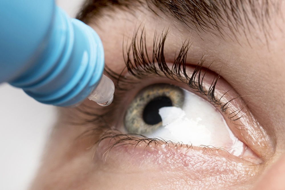
Adult Nasolacrimal Duct Obstruction

Nasolacrimal duct obstruction prevents tears from draining into the nose, causing persistent watering, discharge and recurrent infection. In adults it most often occurs where the duct enters the nose, and complete obstruction is treated surgically by creating a new passage between the lacrimal sac and the nose.

Congenital Nasolacrimal Duct Obstruction

The commonest reason a baby's eye waters is that the thin membrane where the tear duct opens into the nose has not opened after birth. The great majority of these congenital blockages resolve within the first twelve months. Lacrimal sac massage, lid hygiene and regular review carry that period, while pictures such as sac infection are assessed without waiting.

What Is the Lacrimal System and How Does It Work?

The lacrimal system has two parts. The secretory part consists of the lacrimal gland, which sits in the upper outer corner of the orbit, together with the accessory glands in the lid margin and conjunctiva. The drainage part is the channel system that carries tears from the ocular surface into the nose.

The tear film has three layers: an outer oily layer from the meibomian glands of the lid margin, a middle watery layer, and an inner mucin layer that lets the film adhere to the ocular surface. Every blink renews this film and draws tears into the upper and lower puncta at the inner corner of the eye. From there they pass through the canaliculi, the lacrimal sac and the nasolacrimal duct into the nasal cavity. This is why the nose runs when we cry.

A disorder anywhere along this chain is described as a lacrimal system disorder. Problems on the secretory side cause dryness; problems on the drainage side cause watering.

Why Do the Eyes Water?

Watering (epiphora) is one of the complaints I hear most often. Most patients assume the only cause is a blocked duct, but there are two distinct mechanisms.

Inadequate drainage: Tears are produced normally but cannot drain away. Punctal stenosis, canalicular obstruction, lacrimal sac disease and nasolacrimal duct obstruction belong to this group.

Excess production (reflex tearing): The ocular surface is irritated, so tear production increases. Dry eye, blepharitis, allergy, ingrown lashes, in-turning or out-turning of the lid and foreign bodies belong here.

It may sound contradictory, but dry eye also causes watering. When the surface dries out, a reflex produces dilute, poor-quality tears, and the patient describes both burning and watering. Treatment given without making this distinction does not work. See the dry eye disease and ingrown eyelashes pages for detail.

How Is a Blocked Tear Duct Recognised?

Constant watering that runs down the cheek and worsens in wind and cold.

Crusting that is marked in the morning, with the lashes sticking together.

Swelling beside the bridge of the nose, below the inner end of the lower lid. This may be a distended lacrimal sac.

Purulent material coming back through the punctum when this swelling is pressed.

Repeated episodes of conjunctivitis and frequent redness of the lids.

Sudden pain, redness and swelling spreading towards the cheek. This is acute dacryocystitis and should be assessed without delay.

What Is Dacryocystitis?

Dacryocystitis is inflammation of the lacrimal sac, and there is almost always an underlying duct obstruction. Tears stagnating in the sac create a favourable environment for bacterial growth.

Acute dacryocystitis begins abruptly, with a painful, red, warm swelling beside the bridge of the nose, sometimes with fever and malaise. The infection has to be brought under control first. Chronic dacryocystitis is more insidious: persistent watering, morning crusting and discharge that appears when the sac is pressed.

Settling the infection is only a temporary solution; unless the obstruction is relieved, the picture recurs. Definitive treatment is surgical, and it is planned once the acute episode has settled. See adult nasolacrimal duct obstruction.

In the short video below I describe the newborn form of sac distension, the dacryocystocele.

Which Tests Are Used?

Slit-lamp examination: The lid margin, the position and patency of the puncta, the height of the tear meniscus and the ocular surface are assessed.

Dye disappearance test: Fluorescein is instilled. If most of the dye is still present after five minutes, drainage is inadequate. It is particularly useful in infants and children.

Syringing through the punctum: Saline is injected through a fine cannula. Fluid reaching the throat indicates a patent system; reflux indicates obstruction, and the pattern shows at which level.

Probing: A fine probe identifies the point at which the passage is blocked.

Schirmer test and tear break-up time: These measure tear production and film quality, distinguishing watering caused by dry eye.

Dacryocystography and dacryoscintigraphy: Used to image the drainage system and follow the flow of tears.

CT or MRI: Requested where there is trauma, suspicion of a mass, or sinus disease.

Nasal endoscopy: Allows assessment of the nasal anatomy and of the area where the duct opens into the nose.

Treatment Options

Treatment follows the diagnosis; the same approach is not applied to every kind of watering.

Ocular surface treatment: Where watering is caused by dryness, blepharitis or allergy, the surface is restored with warm compresses, lid hygiene and artificial tears.

Correcting lid position: If the lid is turned outwards (ectropion) or inwards (entropion), the punctum cannot collect tears. Lid surgery often resolves the watering on its own.

Punctal dilation: A narrowed punctum can be widened with a minor procedure.

Silicone tube intubation: In partial narrowing, a fine silicone tube keeps the passage open and is removed after a few months.

Dacryocystorhinostomy (DCR): The operation used for complete obstruction. A new passage is created between the lacrimal sac and the nasal cavity, either through the skin (external) or through the nose (endoscopic).

Conjunctivodacryocystorhinostomy (CDCR): Where the canaliculi cannot be used, a glass tube (Jones tube) is placed between the eye and the nose.

Sac massage and probing in infants: Most congenital obstructions open spontaneously during the first year; probing is used for those that do not.

Options are not exhausted in patients whose symptoms persist after earlier surgery. I have set this out on the revisional surgery for failed nasolacrimal duct obstruction treatment page.

Pages in This Section

Adult nasolacrimal duct obstruction — causes, symptoms, DCR surgery, risks and recovery.

Congenital nasolacrimal duct obstruction — watering in infants, sac massage, probing and intubation.

Revisional surgery after failed tear duct surgery — why surgery fails and what can be done.

Dry eye disease — reduced tear production and impaired tear quality.

Practical Advice

Keep up lid hygiene; the oil glands of the lid margin determine the stability of the tear film.

Blink rate falls in front of a screen. Look into the distance at regular intervals and rest your eyes.

Wear sunglasses to protect your eyes from wind and ultraviolet light.

Do not neglect eye infections; repeated infections can leave permanent narrowing in the drainage system.

Keep nasal conditions such as chronic sinusitis and allergic rhinitis under control.

Do not use eye drops long term on your own initiative; prolonged use of some drops can narrow the puncta and canaliculi.

See an ophthalmologist if watering, discharge, or swelling and pain beside the bridge of the nose persist.

Lacrimal system disorders can largely be brought under control with early diagnosis and appropriate treatment. Identifying the true source of the watering is the step that determines everything that follows.

Ayşe Dolar Bilge MD, FEBOphth.
Ophthalmologist, Oculoplastic Surgery

More than 1000+ happy health support.
10+
Support from healthcare personnel
4.9
200+ Rating
Scroll
CONTACT US
WHATSAPP