
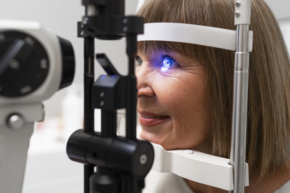
Prosthetic eye users may develop irritation, heavy discharge, infection, a prosthesis that no longer fits, socket contraction and hollowing of the upper lid over time. These affect daily comfort and the fit of the prosthesis, and regular care with an annual review largely prevents them.

Evisceration and enucleation are two different operations performed on an eye that has no vision. Evisceration empties the eye and preserves its outer shell; enucleation removes the globe intact. In both, an implant is placed in the socket and a prosthetic eye is fitted once healing is complete.
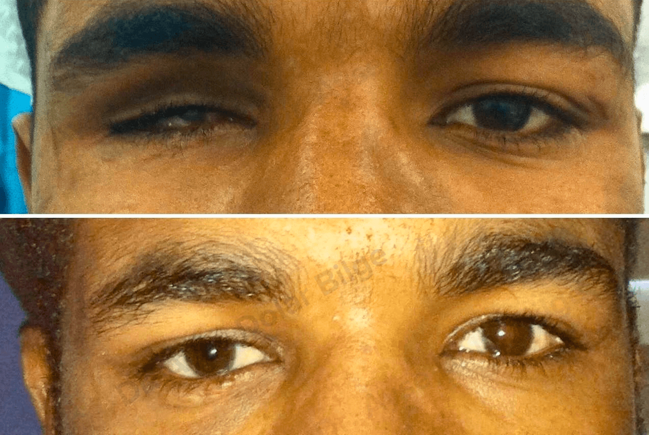
A prosthetic eye, also called an ocular prosthesis, is a custom-made artificial eye placed in the socket of a person born without a developed eye or who has lost an eye to disease, tumour or injury. Its purpose is to restore the appearance of the eye region and to preserve the structure of the socket and eyelids. The first thing I explain to my patients is that a prosthetic eye is a shell, not an organ. It does not replace the eye itself, but it restores facial symmetry and much of the eyelid movement.
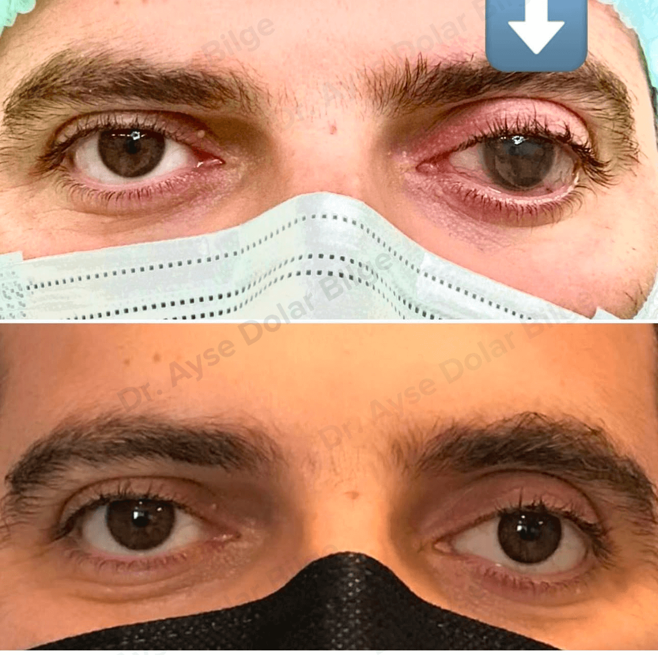
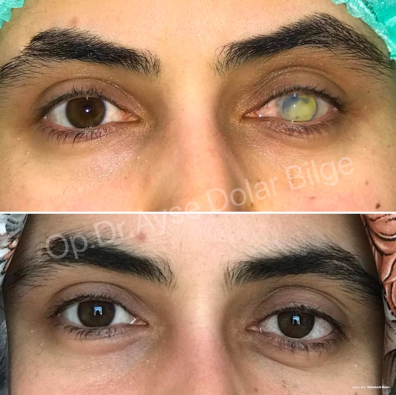
Modern prostheses are not spheres. They are thin, concave acrylic shells that rest on the tissue of the socket and sit behind the eyelids. The colour, the vessel pattern and the iris diameter are hand painted to match the companion eye. A well-made prosthesis therefore looks natural enough that most people facing the patient do not notice it.
No. This is one of the questions I am asked most often, and the answer is clear: a prosthetic eye does not provide vision. Sight depends on light reaching the retina and travelling along the optic nerve to the brain. A prosthesis contains none of these structures — no retina, no lens, no optic nerve.
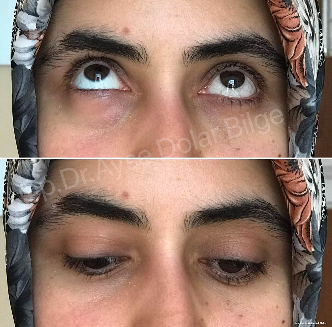
The benefit is structural and aesthetic. The prosthesis maintains the volume of the socket, prevents the lids from collapsing inward, helps tears spread evenly along the lid margin and allows the patient to feel comfortable in daily life. Adapting to seeing with one eye is a separate process. Depth perception is reduced in the first months, and the brain gradually compensates by relying on shadow, size and motion cues. During this period I advise extra care when driving, using stairs or pouring liquids.
The socket must be able to support the prosthesis. If there is marked lid shortening, socket contraction or significant tissue loss, these problems are corrected first. I have described them in detail on the socket problems page.
These three terms are often confused. Evisceration and enucleation are the names of operations; the prosthetic eye is the name of the part fitted afterwards.
Which operation is chosen depends on the condition of the eye. A side-by-side comparison is given on the evisceration and enucleation page.
Immediately after surgery a temporary clear or lightly tinted piece called a conformer is placed in the socket. Its role is to keep the lids from sticking together and to stop the socket from contracting. Once the swelling settles and the tissues stabilise, the impression for the definitive prosthesis is usually taken within four to eight weeks.
The definitive prosthesis is made from a mould of the socket, and the colour and vessel pattern are painted with the companion eye in front of the ocularist. Several fine adjustments after the first fitting are normal and are not a sign that something has gone wrong. During the same period we work together on inserting and removing the prosthesis, cleaning it, and recognising the symptoms that require review.
Caring for a prosthesis is easy to learn and important to keep up. This is the routine I recommend:
Opinions differ on how often the prosthesis should be taken out. Because frequent handling can irritate the socket, removing and cleaning it once a week is enough for most patients who have no complaints. Sleeping with the prosthesis in place is not a problem.
Discharge is the most common complaint among prosthesis wearers and on its own it is not always a sign of a serious problem. The usual causes are:
If the discharge turns yellow or green, or if there is pain, marked redness, swelling or bleeding in the socket, the socket should be examined. Further detail is on the socket problems page.
Acrylic prostheses wear down over the years: the surface becomes scratched and the paint fades. In adults, replacing the prosthesis roughly every four to six years, with polishing in between, is usually appropriate. Because the volume of the socket changes over time, the thickness of the prosthesis may also need adjusting.
I recommend a socket and prosthesis check once a year. At that visit we assess the fit of the prosthesis, the position of the lids, the depth of the fornices and the health of the lining. Correcting a small misfit early is far easier than treating an advanced socket problem.
The situation in children differs from that in adults. The eyeball acts as a natural spacer that stimulates the bony growth of the orbit. If the eye is lost early or never develops, the socket and that half of the face can fall behind in growth.
For this reason a conformer is fitted as early as possible in infants and young children, and the socket is then expanded gradually with progressively larger prostheses. Depending on the rate of growth, changes are needed anywhere between every few months and once a year. Teaching the family to insert and remove the prosthesis, and keeping to the review schedule, makes the difference in this period.
With sound surgery and regular follow-up, a prosthetic eye can be worn comfortably for many years. If you notice a change in your socket, it is worth having it assessed rather than waiting.
Ayşe Dolar Bilge MD, FEBOphth.
Ophthalmologist, Oculoplastic Surgery
 English
