Birgen Business Center, Bağdat Ave. No 226, Floor 2, Unit 10, Kadıköy, Istanbul, Turkey
+90 553 910 91 39 info@aysedolarbilge.com
Contact Us WHATSAPP
Get Information Now
Please enter a valid name.
Lütfen geçerli bir Email adresi giriniz.
Please enter a valid phone number.
Prosthetic eye users may develop irritation, heavy discharge, infection, a prosthesis that no longer fits, socket contraction and hollowing of the upper lid over time. These affect daily comfort and the fit of the prosthesis, and regular care with an annual review largely prevents them.
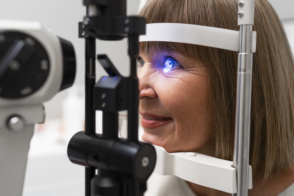
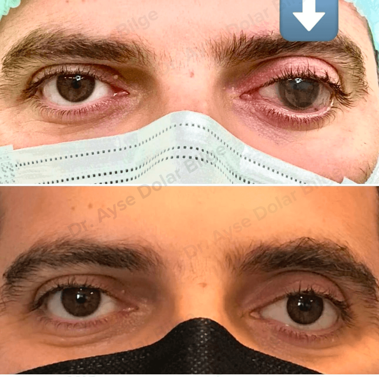
Clinical assessment of a prosthetic eye socket

Prosthetic eye users may encounter a range of socket problems over time. These affect both daily comfort and the appearance and fit of the prosthesis. Most can be prevented with regular care and timely review, and when they do occur they are straightforward to deal with if recognised early.

What Is the Prosthetic Eye Socket?

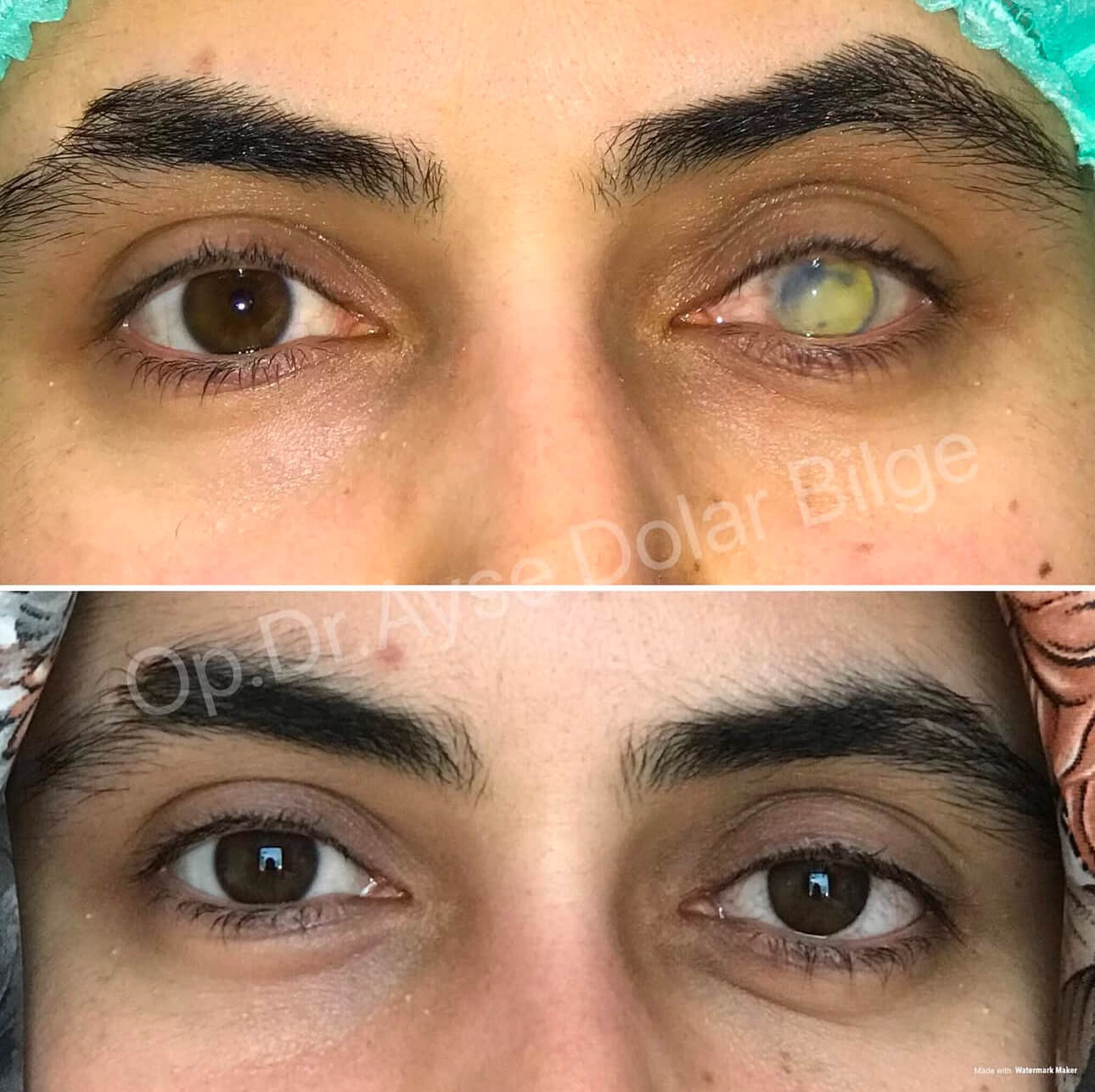
The socket is the space remaining after evisceration or enucleation, lined with conjunctiva. Its depth, the position of the lids and the volume it retains all determine how the prosthesis will sit.

The orbital implant placed at surgery provides most of the volume. The prosthesis is a thin shell that sits in front of the implant, behind the eyelids. Socket, lids and prosthesis form a connected balance, and a change in one affects the others. For the operations themselves see the evisceration and enucleation page.

Common Socket Problems

Irritation and Discomfort

The socket may feel gritty, itchy or burning.

Reduced tear production or poor tear quality causes dryness and discomfort.

Scratches and roughness on the prosthesis irritate the lid tissue.

Excessive Mucus and Discharge

The socket lining can produce heavy mucus as a reaction to the prosthesis.

Mucus build-up makes the prosthesis look cloudy and increases the need for cleaning.

Discharge that turns yellow or green, with accompanying pain, suggests infection.

Infection and Inflammation

Redness, discharge, swelling and pain may indicate infection.

Poor hygiene and irregular cleaning of the prosthesis increase the risk.

Chronic inflammation of the socket lining can cause papillary changes on the surface.

A Prosthesis That No Longer Fits

Over time the prosthesis may not seat properly, creating pressure points, pain and tissue damage.

A worn surface reduces comfort and increases mucus production.

A poorly fitting prosthesis may fall out repeatedly or fail to stay in place.

Socket Contraction

The socket can shrink because of insufficient conjunctiva or scar tissue.

When the fornices between lid and socket become shallow, the prosthesis cannot stay in place.

Leaving the socket empty for long periods accelerates contraction, which is why a prosthesis or conformer should stay in place continuously.

Volume Loss and Upper Lid Hollowing

Soft tissue volume in the socket can decrease over the years.

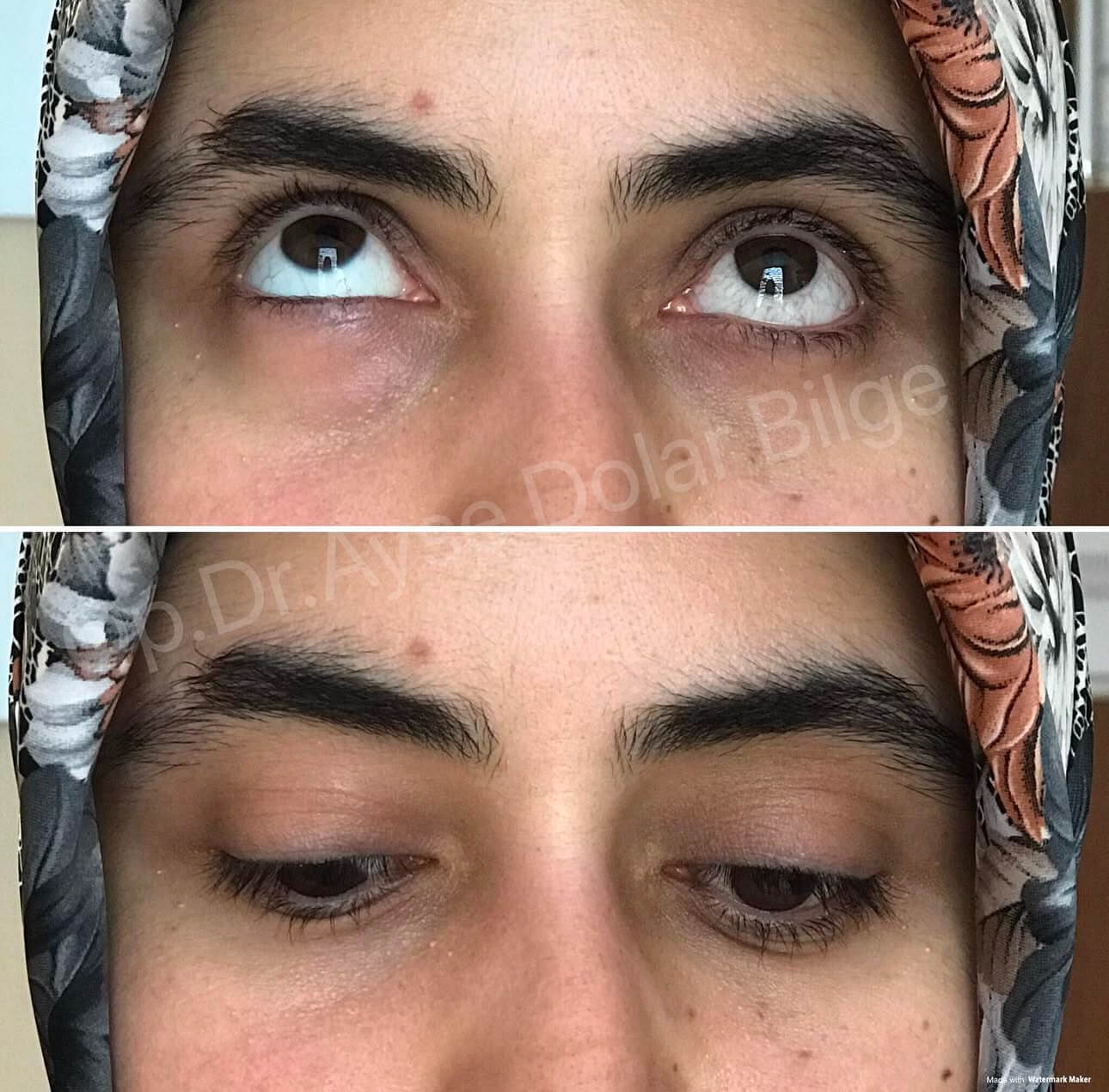
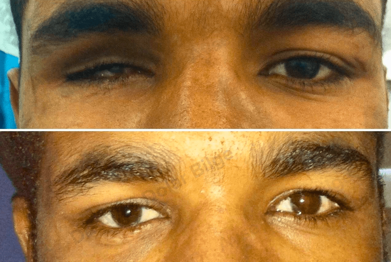
A marked hollow above the upper lid, lid droop and laxity of the lower lid may develop.

This picture is called anophthalmic socket syndrome and noticeably affects the appearance of the prosthesis.

Lid Position Problems

The lower lid can become lax and sag, so that it no longer supports the prosthesis.

The lid may turn inwards (entropion) or outwards (ectropion).

Drooping of the upper lid can cover part of the prosthesis.

Implant-Related Problems

A whitish area appearing in the socket may indicate that the tissue over the implant has opened (exposure).

The implant can move or change position within the socket.

These findings should be assessed without delay.

Why Do These Problems Develop?

Irregular cleaning of the prosthesis and inattention to hand hygiene.

Cleaning with abrasive products such as alcohol, cologne, bleach or toothpaste.

A prosthesis left unchanged for many years, with a worn surface.

Changes in socket volume over time.

Reduced tear production, blepharitis and allergy.

Leaving the prosthesis untouched for long periods, or conversely handling it far more often than needed.

A history of radiotherapy or severe trauma.

Missed follow-up appointments.

How Is the Socket Assessed?

Examination with the prosthesis in place: Its position, the symmetry of the lid opening and its participation in eye movement are assessed.

Inspection of the prosthesis: Surface scratches, worn edges, deposits and colour match are checked.

Socket examination: With the prosthesis removed, the health of the lining, the depth of the fornices, scar tissue and the tissue over the implant are inspected.

Lid assessment: Lower lid laxity, upper lid position and closure are measured.

Tear assessment: Where dryness is a complaint, tear production and the lid margin are examined.

Imaging: CT or MRI is requested where there is a suspected implant problem or a mass in the socket.

Examination of the remaining eye: Carried out at every visit; protecting the seeing eye is the priority.

Solutions and Treatment Options

Regular hygiene and care: Cleaning the prosthesis at appropriate intervals, washing hands before handling it and avoiding abrasive products.

Polishing the prosthesis: Polishing a dulled surface markedly reduces mucus production and irritation.

Replacing the prosthesis: A worn prosthesis, or one that no longer fits, is replaced. In adults, replacement roughly every four to six years is appropriate.

Adjusting the thickness: Where volume loss is mild, building up the back surface of the prosthesis can compensate.

Artificial tears: Preservative-free artificial tears can be used on medical advice for dryness and comfort.

Lid margin care: Warm compresses and lid hygiene where there is blepharitis.

Treating infection: Where there is redness, pain and purulent discharge, appropriate treatment is arranged after assessment.

Lid surgery: Lower lid laxity, lid droop and in- or out-turning are corrected surgically.

Socket expansion and grafting: In advanced contraction, mucous membrane or tissue grafts can be used to deepen the fornices.

Implant procedures: An exposed implant is covered and, where necessary, exchanged; additional volume can be provided where there is deficiency.

Preventing Socket Problems

Always wash your hands with soap before handling the prosthesis.

Clean it over a sink with a towel laid underneath.

Do not use alcohol, cologne, bleach, toothpaste or stiff brushes.

Do not rub the socket; wipe the lid margin with moistened clean cotton, from the inner corner outwards.

Do not leave the prosthesis or conformer out for long periods, as the socket contracts.

Have the socket and prosthesis reviewed at least once a year.

In children, review intervals are much shorter because growth continues.

Protect your remaining eye, and use protective eyewear for hazardous work and sports.

When Should You Seek Medical Advice?

Persistent pain, throbbing or a feeling of pressure in the socket.

Increasing redness, swelling, warmth, or dark-coloured and foul-smelling discharge.

A prosthesis that repeatedly falls out, will not stay in place or cannot be inserted.

A whitish area appearing in the socket.

A deepening hollow above the upper lid, or a lid that no longer closes fully.

Bleeding from the socket.

Blurring, flashes of light or reduced vision in the remaining eye.

Socket problems can largely be prevented with regular care and specialist review. If you feel any discomfort, it is worth having it assessed rather than waiting. For prosthesis care and replacement intervals, see the prosthetic eye page.

Ayşe Dolar Bilge MD, FEBOphth.
Ophthalmologist, Oculoplastic Surgery
Get Online Consultation

As an Ophthalmologist, Dr. Ayşe Dolar Bilge, I provide diagnosis and treatment services with my experienced team.

Frequently Asked Questions

Why does a prosthetic eye produce discharge?

The most common reason is the surface of the prosthesis becoming dull with deposits building up on it; polishing usually resolves this. Reduced tear production or poor tear quality also makes the socket produce heavy mucus in response. A prosthesis that no longer fits, inflammation of the lid margin and lapses in the cleaning routine are other causes. Discharge that turns yellow or green, with pain, should be assessed.

How is a prosthetic eye cleaned?

Wash your hands with soap and work over a sink with a towel laid underneath. Rinse the prosthesis with lukewarm water; if an oily film remains, wash it with a neutral, unscented soap and rinse thoroughly. Do not use alcohol, cologne, bleach, toothpaste or stiff brushes, as these damage the polished surface. Do not rub the socket; wipe crusting from the lid margin with moistened clean cotton, from the inner corner outwards.

How often should a prosthetic eye be replaced?

Acrylic prostheses wear down over the years: the surface becomes scratched and the paint fades. In adults, replacement roughly every four to six years with polishing in between is usually appropriate. Because socket volume changes over time, the thickness may also need adjusting. In children, replacement is needed far more often as the facial bones grow. I recommend a socket and prosthesis check once a year.

My prosthesis keeps falling out — why?

The most common reasons are a prosthesis that no longer fits the socket, a lax lower lid that cannot support it, and shallowing of the fornices. A worn prosthesis whose edges have lost their shape also holds less well. The fit of the prosthesis, the position of the lids and the depth of the socket are assessed together. The answer is sometimes a new prosthesis and sometimes surgical correction of lid laxity.

What causes socket contraction and how is it prevented?

Socket contraction can result from insufficient conjunctiva, scar tissue, previous radiotherapy or severe trauma. The most common preventable cause is leaving the socket empty for long periods; once the prosthesis or conformer is removed, the socket begins to shrink quickly. The prosthesis should therefore stay in place continuously. In advanced contraction, mucous membrane or tissue grafts can be used to deepen the fornices.

What are the signs of infection in the socket?

Increasing redness, swelling, warmth, persistent pain and dark-coloured or foul-smelling discharge suggest infection. Bleeding from the socket also needs assessment. With these signs, do not force the prosthesis in or out, and seek advice without delay. A whitish area appearing in the socket may indicate that the tissue over the implant has opened, which should also be assessed promptly.

Can hollowing of the upper lid be corrected?

Hollowing of the upper lid results from volume loss in the socket over time and is part of anophthalmic socket syndrome. Where the loss is mild, building up the back surface of the prosthesis can compensate. In more marked cases, adding volume to the socket or surgically correcting accompanying lid droop is considered. The appropriate method is chosen by assessing socket depth and lid position together.

To get detailed information, send us your contact information and we will call you as soon as possible. Contact us now

Open WhatsApp
Scroll
CONTACT US
WHATSAPP