

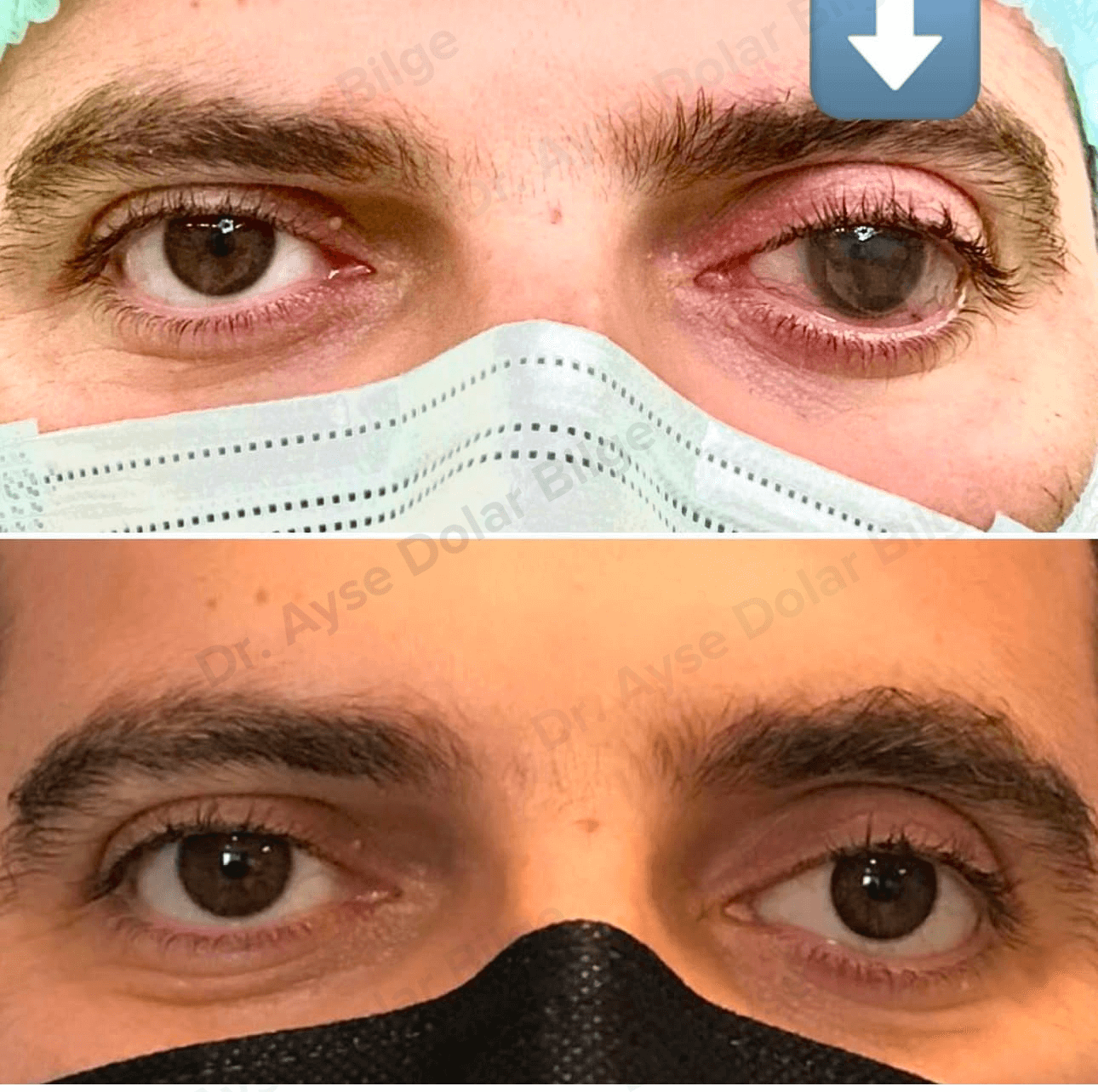
Evisceration and enucleation are two different surgical procedures performed in ophthalmology. They are considered in cases of eye trauma, intraocular tumours, severe infection or a painful blind eye. Both are carried out on an eye that has no vision, and a prosthetic eye is fitted afterwards to preserve facial symmetry.
Evisceration is a surgical procedure in which the internal structures of the eyeball are removed while the sclera — the white outer coat of the eye — and the extraocular muscles attached to it are left in place. In short, the eye is emptied and its outer shell is preserved.
A round orbital implant is placed in the space created, to maintain the volume of the socket, and the sclera is closed over it. Because the muscles remain attached to the sclera, the implant and the prosthesis fitted over it share in the movement of the socket.
Enucleation is a surgical procedure in which the entire eyeball, including the sclera, is removed. The extraocular muscles are detached from the globe and reattached to an orbital implant placed in the socket. The optic nerve is divided behind the globe.
Because the intraocular tissues are removed intact, pathological examination can be carried out completely. In tumour cases this is decisive.
The fundamental difference is whether the outer shell of the eye is preserved.
The decision takes account of the condition of the eye, the underlying disease, the patient's general health and the surgical assessment together.
Some of these appear only later and are uncommon, and most can be corrected when recognised early. For detail see the socket problems page.
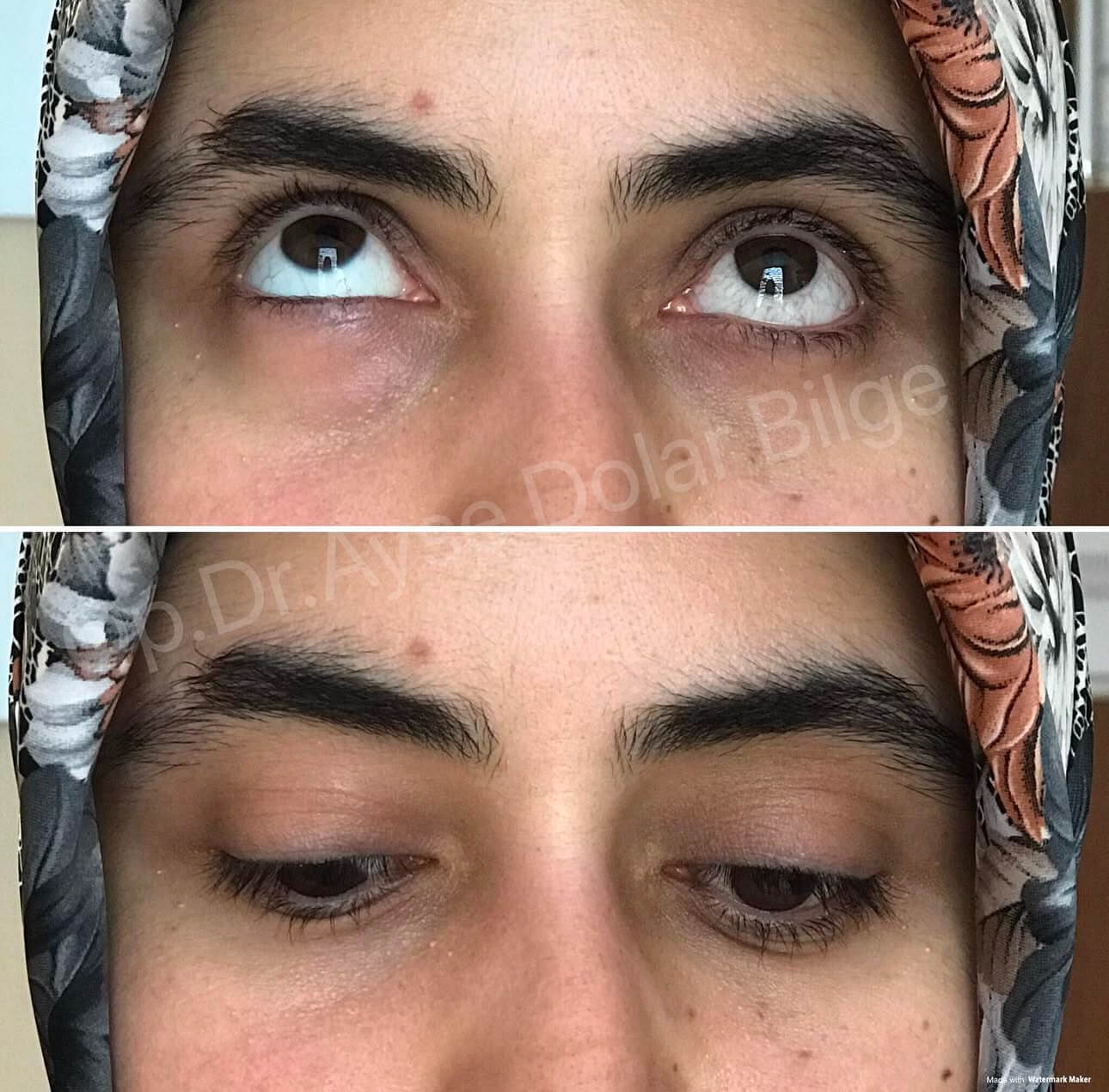
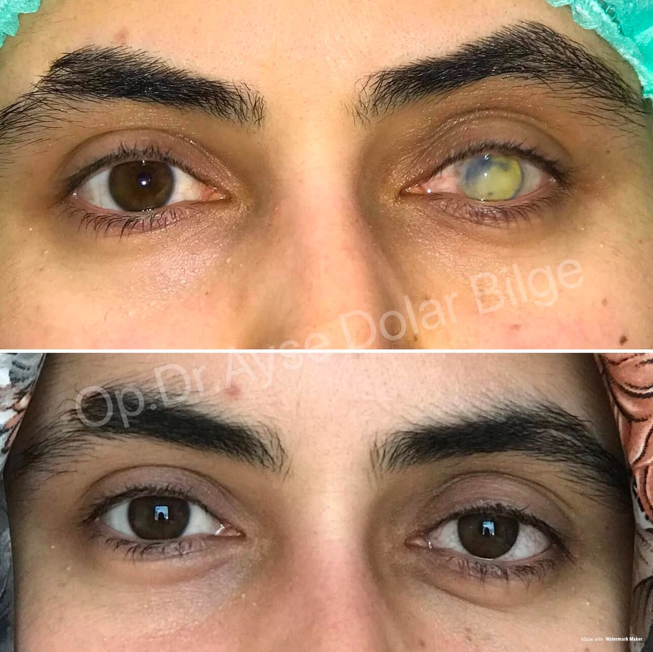
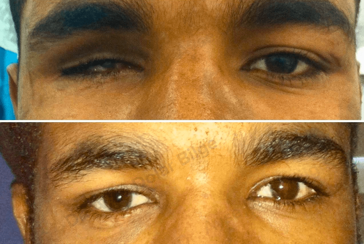
Both operations provide a suitable foundation for a prosthetic eye. The conformer fitted immediately after surgery is temporary; the definitive prosthesis is made individually once the swelling has settled and the tissues have stabilised. Its colour and vessel pattern are hand painted to match the companion eye.
A prosthetic eye does not restore sight. It maintains the volume of the socket, prevents the lids from collapsing and gives a natural appearance. For how it is cleaned, how often it is replaced and how it is planned in children, see the prosthetic eye page.
Evisceration and enucleation are procedures chosen for specific clinical situations, and their outcomes are managed well with regular follow-up. Both provide a foundation for a prosthetic eye, and the aim throughout is to protect the patient's comfort in daily life.
Ayşe Dolar Bilge MD, FEBOphth.
Ophthalmologist, Oculoplastic Surgery
As an Ophthalmologist, Dr. Ayşe Dolar Bilge, I provide diagnosis and treatment services with my experienced team.
Evisceration is an operation in which the internal structures of the eyeball are removed while the sclera — the white outer coat — and the eye muscles attached to it are left in place. A round implant is placed in the space created, to maintain the volume of the socket. Because the muscles stay attached to the sclera, movement of the prosthesis fitted afterwards is generally more natural.
Enucleation is an operation in which the entire eyeball, including the sclera, is removed. The eye muscles are detached from the globe and reattached to an orbital implant placed in the socket, and the optic nerve is divided behind the globe. Because the tissues are removed intact, pathological examination can be carried out completely, which is decisive in intraocular tumour cases.
The fundamental difference is whether the outer shell of the eye is preserved. Evisceration removes the contents and leaves the sclera with the muscles attached to it. Enucleation removes the globe intact and the muscles are reattached to the implant. Prosthetic movement is therefore usually better after evisceration, and recovery is faster. Where there is an intraocular tumour, enucleation is used and evisceration is not appropriate.
The main situations are eye trauma beyond repair, a blind and painful eye that does not respond to medication, severe intraocular infection that has not responded to treatment, intraocular tumours, and a markedly shrunken eye. The decision takes account of the state of the eye, the underlying disease, the patient's general health and the surgical assessment together.
A temporary clear conformer is placed in the socket at the end of surgery, to keep the lids from adhering and the socket from contracting. Once the swelling has settled and the tissues have stabilised, the impression for the definitive prosthesis is usually taken within four to eight weeks. The definitive prosthesis is custom made, with the colour and vessel pattern hand painted to match the companion eye.
Swelling, bruising and a dull ache around the eye are usual in the first days; cold application and keeping the head elevated help. The drops or ointments prescribed are used regularly. Swelling settles noticeably within two to four weeks. During this period heavy lifting, swimming pools and dusty environments should be avoided. The impression for the definitive prosthesis is taken once the tissues have stabilised.
Possible problems include bleeding and haematoma within the socket, infection, exposure or displacement of the implant, contraction of the socket due to insufficient conjunctiva, hollowing of the upper lid and lid droop, and a prosthesis that no longer fits because of volume loss. There are also risks related to anaesthesia. Most of these can be corrected when recognised early, which is why regular review matters.
The operation does not directly affect vision in the other eye. Adapting to seeing with one eye is a process, however: depth perception is reduced in the first months, and the brain gradually compensates using shadow, size and motion cues. Care is needed when driving and on stairs during this period. Protecting the remaining eye is the highest priority, so reduced vision, flashes of light or pain should be assessed without delay.
To get detailed information, send us your contact information and we will call you as soon as possible. Contact us now
 English
