Birgen Business Center, Bağdat Ave. No 226, Floor 2, Unit 10, Kadıköy, Istanbul, Turkey
+90 553 910 91 39 info@aysedolarbilge.com
This section covers the main eye conditions of childhood, from refractive errors and strabismus to eyelid problems and tear duct obstruction. Because the visual pathways are shaped in the first years, examination on schedule is the most effective way to prevent amblyopia.

Eyelid Infections in Children

Eyelid infections in children include styes, chalazia, blepharitis and cellulitis. Most are mild and settle with warm compresses and drops. When fever, forward displacement of the eye or restricted eye movements appear, the infection may have reached the orbit and needs urgent care.
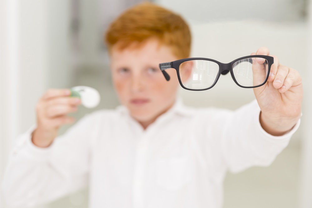
Astigmatism in Children

Astigmatism is a refractive error caused by irregular curvature of the cornea or lens, so light cannot focus at a single point on the retina. In children it causes blurred vision, squinting, headaches and reading difficulty, and untreated it can lead to amblyopia. Early examination protects visual development.
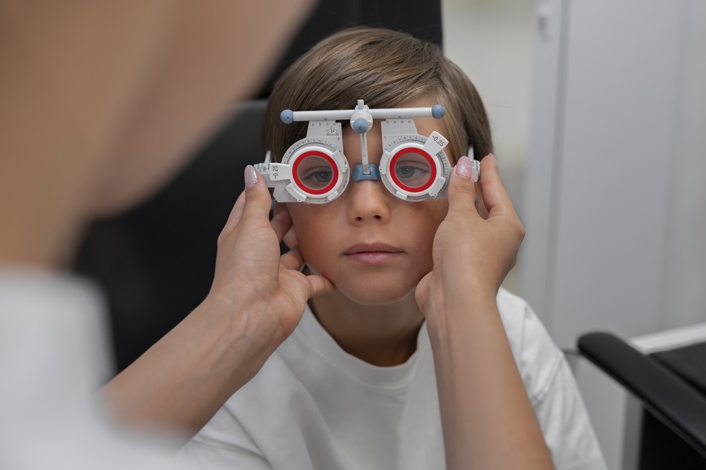
Hyperopia in Children

Hyperopia is a refractive error in which light focuses behind the retina. Children can read the chart normally yet struggle with near work, develop headaches or show an inward turning eye. Cycloplegic refraction reveals the true degree, and the right glasses prevent esotropia and amblyopia.
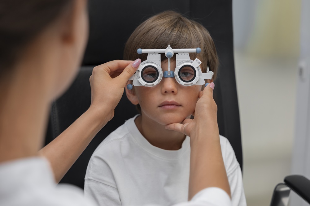
Myopia in Children

Myopia makes distant objects blurry because light focuses in front of the retina. It usually starts at school age and can increase during growth. Trouble seeing the board, squinting and sitting close to the television are the first signs. Correct glasses and regular follow-up protect vision and track progression.
Strabismus in Children

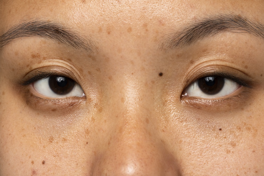
Strabismus means the eyes cannot fix on the same point. Because a child's brain suppresses the deviating eye, untreated squint leads to amblyopia. Glasses, patching and, when needed, muscle surgery can restore both alignment and vision, and the age at which treatment starts shapes the result.
Child Glaucoma

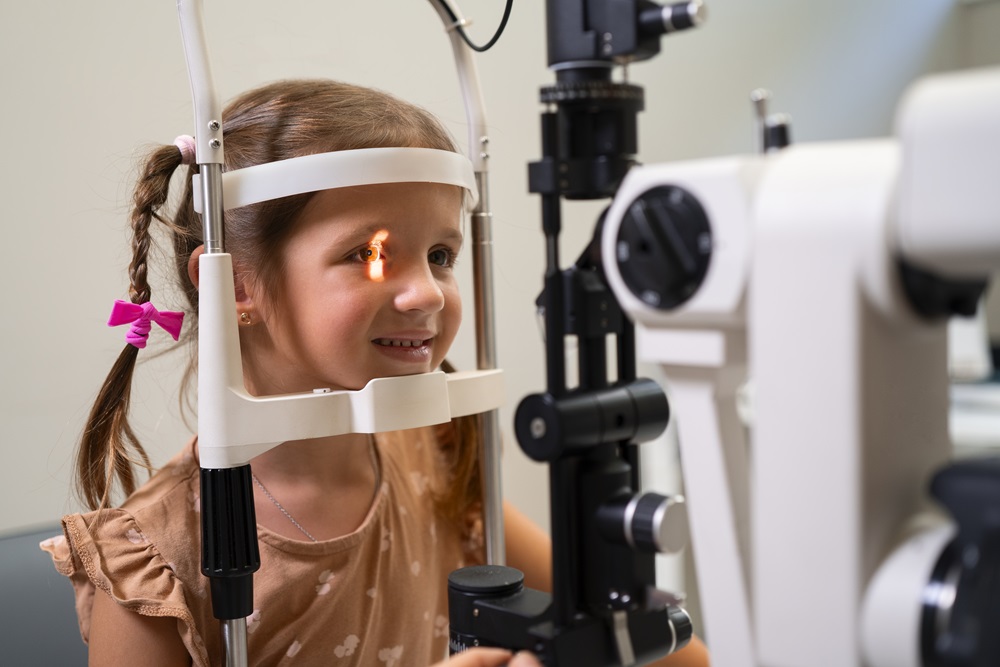
Childhood glaucoma is raised intraocular pressure that damages the optic nerve. Because an infant's eye is elastic, it enlarges under pressure, so large eyes, light sensitivity and constant watering are the key warning signs. Surgery is the main treatment and follow-up continues for life.
Congenital Cataracts

Congenital cataract is clouding of the lens present at birth or appearing in the first months. A white pupil, failure to follow objects and nystagmus are the main signs. Because it occurs while the visual system is developing, the timing of surgery directly determines the outcome.

Child Structural Disorders of the Eyelid

Structural eyelid disorders in children include ptosis, entropion, ectropion, epiblepharon, coloboma and eyelid masses. When the lid cannot protect the cornea or blocks the visual axis, irritation, corneal damage and amblyopia can follow, and treatment is planned around those risks.
Child Eyelid Drooping

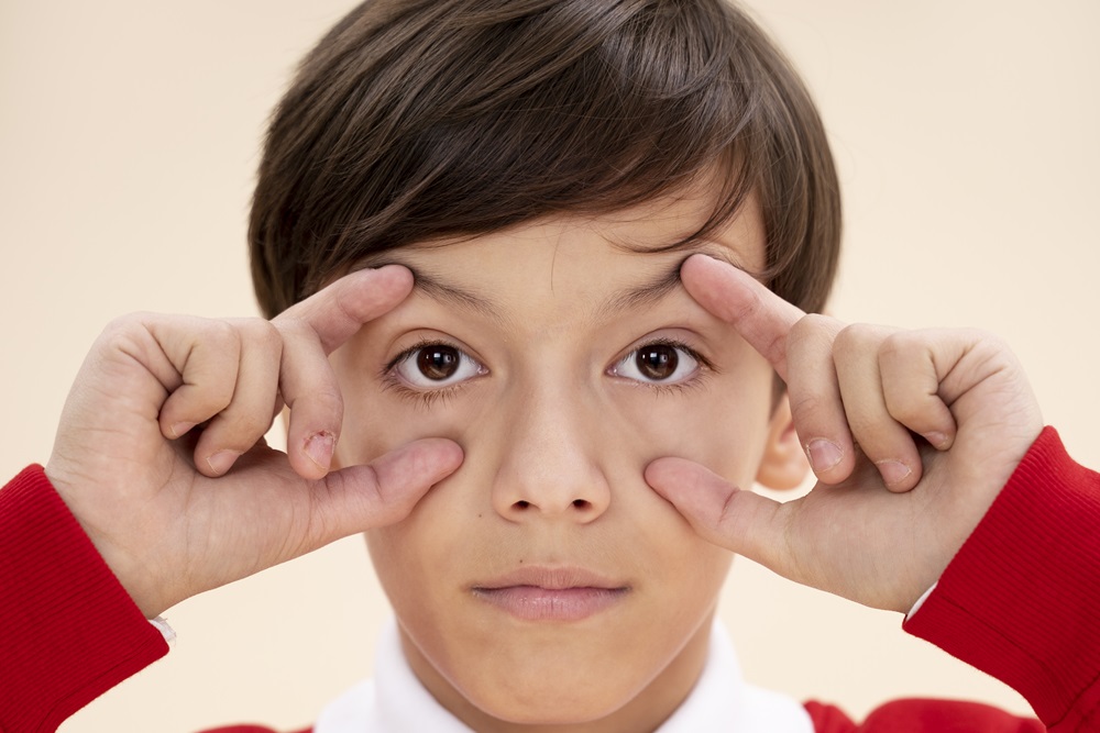
Ptosis in children means the upper eyelid sits lower than normal. When the lid covers the pupil, or presses on the cornea and induces astigmatism, amblyopia follows. The decision to treat is therefore based on visual development rather than on appearance.

Eyelid Vascular Masses (Hemangiomas)

Eyelid haemangiomas are common benign vascular lesions of infancy. They grow rapidly in the first months and then shrink over years. Lesions that pull the eyelid down or press on the cornea can cause amblyopia and are treated rather than watched.
Congenital Mass or Cysts of the Eyelid and Around the Eyes

Most masses and cysts found around a child's eyelid at birth or in the first months are benign. Dermoid cysts, haemangiomas, epibulbar dermoids and lacrimal cysts are the commonest. What guides management is the effect on the eyelid and on vision, not the appearance.

Child Tear Duct Obstructions

When a congenital tear duct blockage has not opened by the first birthday, intervention comes onto the agenda. Probing, silicone intubation, balloon dacryoplasty and, rarely, duct surgery are used from that point. This page covers timing, how each procedure is carried out, what is done when the blockage recurs, and long-term follow-up.

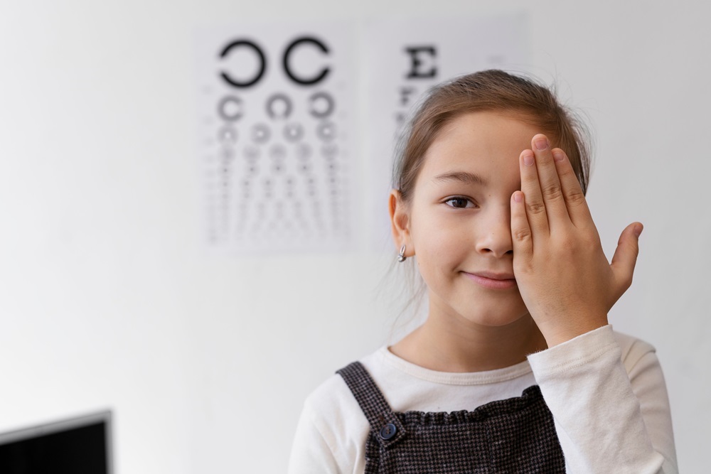
Why Children's Eye Examinations Should Not Be Postponed

Sight is not a ready-made ability. A baby's eye is anatomically complete at birth, but the brain has not yet learned to see. The sharp image falling on the retina shapes the visual pathways through the first years. If one eye cannot receive a clear image during that period, its development falls behind and amblyopia (lazy eye) sets in.

The hardest part of amblyopia is how quietly it progresses. The child copes with the better eye, reads, plays, and never complains. Parents notice nothing, because there is nothing to notice. The problem usually surfaces only at a screening examination. Since the visual pathways are most adaptable in the preschool years, what matters most in paediatric ophthalmology is not a symptom the family reports but an examination performed on time.

Eye Examination Schedule by Age

This is the schedule I recommend for a healthy child without complaints. Intervals are shortened where there is a family history of eye disease, premature birth or developmental delay.

Newborn Period

A red reflex examination confirms the clarity of the ocular media.

This simple test detects congenital cataract and other intraocular conditions.

Eyelid structure, palpebral opening and the size of the eye are assessed.

0-6 Months

Whether the baby looks at faces, makes eye contact and follows moving objects is observed.

Constant watering and discharge are assessed; the commonest cause is tear duct obstruction.

Brief, occasional deviations may be normal at this age.

6-12 Months

A first ophthalmic examination belongs here when there is a family history of refractive error, strabismus or amblyopia.

Preterm infants follow a separate surveillance programme.

Any deviation persisting beyond about four months of age, or that is constant, needs assessment.

Around Age 3

The first age at which visual acuity can be measured with picture charts.

I advise a screening examination for every child; in a child without complaints, amblyopia is most often caught at this age.

Each eye is measured separately.

Before Starting School

A full examination including cycloplegic refraction.

The need for glasses, ocular alignment and stereopsis are assessed.

An uncorrected refractive error makes the first year of school unnecessarily hard.

School Age

Every two years without complaints, yearly for children wearing glasses.

Every six months when the prescription is increasing rapidly.

Difficulty seeing the board, avoiding homework and headaches deserve an eye examination before they are put down to inattention.

What Is Checked at Each Age

Infancy: Clarity of the ocular media, size of the eye, lid structure, watering and alignment.

Preschool years: Visual acuity, refractive error, amblyopia and strabismus.

School age: Change in prescription, the rate of myopia progression, and how the two eyes work together.

Any age: Eyelid masses, drooping or distortion; fundus examination and intraocular pressure where there is risk.

Warning Signs Parents Can Notice

A deviating eye, or the light reflex appearing in different positions in the two eyes on photographs.

A white pupil, or a missing red reflex in one eye on flash photographs.

Tilting the head, lifting the chin or covering one eye to look.

Sitting close to the television or holding books very near, and being unable to read the board.

Constant squinting, eye rubbing and discomfort in bright light.

Persistent watering or discharge in a baby, or eyes that look unusually large.

Drooping, swelling or a coloured lump on the eyelid.

Skipping lines while reading, tiring quickly and headaches.

In the short video below I explain what a swelling at the inner corner means in a baby, and when it should not wait.

What the Examination Involves

Visual acuity tests with picture or letter charts appropriate to the child's age, each eye separately.

Assessment of eye movements and alignment using cover tests.

Cycloplegic refraction: Drops relax the focusing muscle so the true prescription can be measured. Spectacle decisions in children rest on this.

Fundus examination and biomicroscopy of the retina, optic nerve and anterior structures.

Intraocular pressure measurement where there is risk or a suggestive finding.

After cycloplegic drops the pupils stay large for a few hours, near vision is blurred and bright light is uncomfortable. It helps to avoid scheduling the visit on a demanding school day and to bring sunglasses or a hat.

Treatment Options

Glasses or Contact Lenses: To correct refractive errors such as myopia, hyperopia and astigmatism.

Patching Therapy: To treat amblyopia by strengthening the weaker eye.

Surgery: For strabismus, eyelid drooping, tear duct obstruction and glaucoma.

Medications: For infections or inflammation.

Observation: For conditions such as haemangiomas and congenital cysts, where the effect on the eye guides the decision.

Topics in This Section

Astigmatism in children — blurred, shadowed vision, squinting and reading difficulty.

Myopia in children — difficulty seeing the board and progression of the prescription.

Hyperopia in children — near-work strain and its link with inward deviation.

Strabismus in children — misalignment, patching and surgery.

Child glaucoma — enlarged eyes, light sensitivity and intraocular pressure.

Congenital cataracts — white pupil and the timing of surgery.

Child eyelid drooping — ptosis and visual development.

Structural disorders of the eyelid — entropion, ectropion, epiblepharon.

Eyelid infections in children — styes, chalazia and lid swelling.

Child tear duct obstructions — watering, massage and probing.

Congenital masses or cysts — dermoid cysts and others.

Eyelid vascular masses — the course and treatment of infantile haemangiomas.

Advice for Parents

Keep to the recommended examination schedule.

A balanced diet supports general eye health.

Limit screen time; one to two hours outdoors daily is particularly valuable against myopia progression.

Seek an examination promptly for redness, watering or any deviation of the eyes.

A correctly prescribed pair of glasses does not weaken a child's eye; it allows it to develop.

Conclusion

Most childhood eye conditions can be treated successfully when they are caught in time. What is usually lost is not the condition itself but the time. Bringing your child for examination at the recommended ages, even without any complaint, is the most practical way to protect visual development.

Ayşe Dolar Bilge MD, FEBOphth.
Ophthalmologist, Oculoplastic Surgery

More than 1000+ happy health support.
10+
Support from healthcare personnel
4.9
200+ Rating
Scroll
CONTACT US
WHATSAPP