
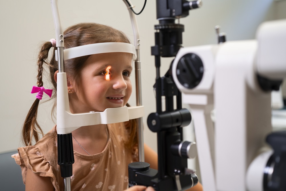
Glaucoma is an eye condition in which raised intraocular pressure damages the optic nerve and can lead to loss of vision. Fluid produced continuously inside the eye normally drains through channels in the anterior chamber angle. When those channels are underdeveloped from birth or become blocked later, fluid accumulates, pressure rises and the optic nerve is harmed.
Childhood glaucoma is far less common than the adult form, but it differs in one important way: the outer wall of an infant's eye is still elastic. Under high pressure the globe expands and the eye looks enlarged. This sign, called buphthalmos, is usually the first thing parents notice, and it is often taken simply for large, striking eyes. Whenever enlarged eyes occur together with light sensitivity and constant watering, examination is essential.
Arises due to other eye conditions, injuries, or systemic illnesses. Examples include:
In a constantly watering infant, the first diagnosis to consider is nasolacrimal duct obstruction, which is far more common. But when watering is accompanied by marked light sensitivity, corneal haze or enlargement of the eye, the picture is different. Measuring corneal diameter and assessing intraocular pressure separates the two. See child tear duct obstructions for the more common condition.
In small infants some of these measurements cannot be obtained reliably while awake, so the examination may be completed under brief anaesthesia. Parents understandably worry about this, but treatment cannot be planned without accurate measurements.
Treatment depends on the type and severity. The key difference from adult glaucoma is that in children, particularly in congenital glaucoma, surgery is the main route. Medication is generally used to bridge the time before surgery or to support control afterwards.
Other topics are listed under pediatric eye diseases.
As an Ophthalmologist, Dr. Ayşe Dolar Bilge, I provide diagnosis and treatment services with my experienced team.
Buphthalmos is enlargement of the eyeball under raised intraocular pressure. Because the outer wall of an infant's eye is still elastic, the globe expands and the eye looks large. When one-sided, the difference between the eyes is obvious. Enlargement together with light sensitivity and constant watering calls for prompt examination.
The commonest cause of constant watering in a baby is nasolacrimal duct obstruction. However, when watering comes with marked light sensitivity, corneal haze or enlargement of the eye, glaucoma must be considered. Corneal diameter measurement and pressure assessment make the distinction.
Unlike in adults, surgery is the main route in children, particularly in congenital glaucoma. Drops are generally used to lower pressure until surgery or to support control afterwards. Expecting long-term control from drops alone is not realistic in most cases.
Measurements such as intraocular pressure, corneal diameter and the drainage angle cannot be obtained reliably in a small infant who is awake, and pressure readings in a crying baby are misleading. The examination is therefore completed under brief anaesthesia when necessary, because treatment cannot be planned without accurate figures.
The aim of surgery is to lower pressure and protect the optic nerve. Final vision depends on more than pressure: corneal haze, high refractive error from elongation of the globe, and amblyopia all play a part. Glasses after surgery and patching where needed are therefore an inseparable part of the process.
Glaucoma is not a condition that is finished once treated. Pressure can rise again years later, usually without symptoms. Follow-up is therefore frequent in the early postoperative period and continues at regular intervals for life. Good vision is not a reason to stop attending.
To get detailed information, send us your contact information and we will call you as soon as possible. Contact us now
 English
