Birgen Business Center, Bağdat Ave. No 226, Floor 2, Unit 10, Kadıköy, Istanbul, Turkey
+90 553 910 91 39 info@aysedolarbilge.com
Eyelid haemangiomas are common benign vascular lesions of infancy. They grow rapidly in the first months and then shrink over years. Lesions that pull the eyelid down or press on the cornea can cause amblyopia and are treated rather than watched.
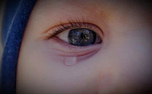
Infant with a red haemangioma on the eyelid

What Are Eyelid Haemangiomas?

Haemangiomas are non-cancerous growths of blood vessels.

They often appear shortly after birth or within the first few weeks of life.

On the eyelid they present as a red or purple mass.

Most shrink and disappear as the child grows, though some require treatment.

Haemangiomas follow a characteristic course, and knowing it eases much of the anxiety: rapid growth in the first months, then a plateau, then slow shrinkage and fading over the following years. The difficulty is that the rapid growth phase coincides exactly with the period in which the visual system is developing. A haemangioma that pulls the lid down or casts a shadow across the visual axis is not one we can wait out.

Symptoms of Eyelid Haemangiomas

Red or purple lump: A raised, soft, spongy mass on the eyelid.

Swelling: The eyelid may appear swollen or asymmetrical.

Vision problems: If the lesion blocks the line of sight, amblyopia can follow.

Eye irritation: The mass may cause redness, tearing or discomfort.

Distortion of the eyelid and change in its shape.

Swelling when the baby cries or strains, a typical feature of its vascular nature.

Bright red on the surface, or a bluish bulge when deeper, depending on the depth of the lesion.

Causes

Abnormal blood vessel growth: The exact cause is unknown, but it involves overgrowth of vascular cells.

Genetic factors: A family history may increase the risk.

Hormonal influences during infancy may play a role.

Risk groups: More frequent in girls, in preterm and low-birth-weight infants, and in multiple pregnancies.

Natural Course: Growth and Involution

Growth phase: Usually rapid in the first months, with visible enlargement over weeks.

Plateau phase: Growth stops and the lesion stays the same size for a period.

Involution phase: Slow shrinkage over years, with fading colour and softening.

After involution, a slight colour difference, laxity or residual tissue may remain.

The question that guides management is whether the eye will be harmed before that natural course completes. If the answer is yes, we do not wait.

Diagnosis

Physical examination: Size, location and impact of the lesion are assessed.

Imaging tests: Ultrasound or MRI to evaluate depth and extent; imaging is essential if the lesion extends into the orbit.

Eye examination: To check for vision problems or amblyopia.

Cycloplegic refraction: Pressure on the cornea induces astigmatism, and measuring it is the most practical way to catch amblyopia risk early.

Assessment of alignment and eye movements.

Multiple cutaneous lesions or extensive midfacial involvement prompt further investigation with the relevant specialties.

Treatment Options

Observation

Small, asymptomatic haemangiomas may not require treatment and can be monitored over time.

Size, lid position, refraction and vision are followed together.

Photographs taken by the family make changes between visits easier to judge.

Medications

Propranolol: A beta-blocker effective in shrinking haemangiomas and often the first-line treatment.

Steroids: Oral or injected steroids may be used to halt growth.

Because treatment requires monitoring of parameters such as heart rate and blood sugar, it is run together with the paediatrician and only under medical supervision.

The effect is more pronounced when treatment begins during the growth phase.

Laser Therapy

Used for superficial haemangiomas to reduce redness and improve appearance.

It can also be considered for residual colour change after involution.

Surgical Removal

Reserved for large lesions that interfere with vision or where other treatments have not succeeded.

Surgical correction may also be planned at an older age for lax skin or residual tissue left after involution.

Amblyopia Treatment

Glasses when the lesion has induced astigmatism.

Patching the stronger eye when one eye has fallen behind.

Amblyopia does not resolve simply because the lesion shrinks; these steps must be carried out separately.

When to Seek Care Without Delay

A lesion pulling the eyelid down or covering the pupil.

Rapid enlargement over weeks.

Forward displacement of the globe, restricted eye movements or a newly appearing deviation.

Ulceration or bleeding of the surface.

Redness, warmth and fever over the lesion.

Multiple haemangiomas on the skin.

Points That Matter in Follow-Up

Early diagnosis improves the outcome of treatment.

Growth of the lesion and its effect on ocular function should be reviewed regularly.

Family involvement matters throughout treatment.

Do not stop vision monitoring because the lesion is shrinking; the real risk is the amblyopia it leaves behind.

Recommendations for Parents

Seek assessment promptly for any red or purple lump on the eyelid.

Even though most haemangiomas shrink on their own, regular follow-up matters because they can affect ocular function.

Follow the treatment plan and attend all follow-up appointments.

Do not apply pressure to the lesion or put any preparation on it.

For other congenital lesions around the eye see congenital masses or cysts, and for lid drooping see child eyelid drooping. All topics are listed under pediatric eye diseases.

Get Online Consultation

As an Ophthalmologist, Dr. Ayşe Dolar Bilge, I provide diagnosis and treatment services with my experienced team.

Frequently Asked Questions

Do eyelid haemangiomas disappear on their own?

The great majority grow in the first months and then shrink and fade over the following years. But the time that natural course takes is exactly the period in which the visual system develops. If the lesion pulls the eyelid down or presses on the cornea, waiting for spontaneous involution is not appropriate.

How does a haemangioma affect vision?

In two ways. If the lesion covers the pupil, no sharp image reaches the retina and amblyopia develops. If it presses on the cornea, it induces astigmatism, creating a marked difference between the eyes that ends the same way. Cycloplegic refraction is therefore a fixed part of follow-up.

Is propranolol treatment safe?

Propranolol is commonly used and is usually the first-line treatment for periocular haemangiomas. Because it requires monitoring of heart rate, blood pressure and blood sugar, it is run together with the paediatrician under medical supervision, and decisions to start and stop rest on that assessment.

When is surgery used for a haemangioma?

Surgery is reserved for lesions threatening vision where medical and laser treatment have not been sufficient. It may also be used at an older age to correct lax skin or residual tissue remaining after involution.

The lump swells when my baby cries. Is that normal?

Yes, this is typical of vascular lesions. Blood flow to the area increases with crying or straining, so the lesion temporarily swells and darkens before returning to its previous state. Regular follow-up and photographic records help separate this from genuine growth.

Does a haemangioma leave a mark after it fades?

After involution, a slight colour difference, visible vessels, laxity or residual tissue may remain, and some of this fades with time. Laser can be considered for persistent colour change and corrective surgery for excess tissue, usually planned at an older age.

To get detailed information, send us your contact information and we will call you as soon as possible. Contact us now

Open WhatsApp
Scroll
CONTACT US
WHATSAPP