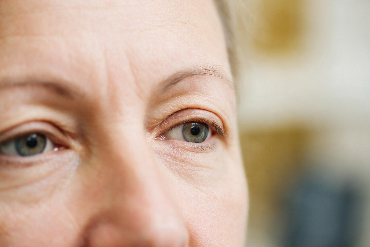
A good number of the people who come to me have already decided what they have: either ptosis or hooded eyelids. About half of them have decided wrong, and the two are not interchangeable. In ptosis the eyelid margin itself sits too low. In hooded eyelids the margin is exactly where it should be and the skin above it has draped forward over it. This article explains what separates the two, what I measure in the clinic, and the situations in which a droopy eyelid is a medical question rather than a cosmetic one.
Ptosis is a low upper eyelid margin. In an eye that opens normally, the upper lid margin covers the top one to two millimetres of the iris. When it covers more than that, the eye looks smaller, the gaze looks tired, and the upper part of the visual field starts to shrink.
The muscle that lifts the lid is the levator. It reaches the tarsus, the firm plate inside the eyelid, through a fan of connective tissue called the aponeurosis. A second, much smaller muscle driven by the sympathetic nervous system adds another millimetre or two of lift.
Ptosis can come from any step of that chain: the muscle, the nerve that drives it, the junction between nerve and muscle, or the aponeurosis pulling away from the tarsus. The surgical side of this is covered on the droopy eyelid (ptosis) page.
Hooded eyelids, described medically as dermatochalasis, are a skin problem rather than a lifting problem. Over the years the upper eyelid skin loses elasticity and comes down over the crease, sometimes as far as the lashes. The lid margin has not moved. The hood sitting on top of it is what makes the eye look closed down.
Brow position confuses this further. When the brow itself descends, it pushes the upper lid skin down and creates the same hooded look without any change in the eyelid. Many people have a mix of all three: a slightly heavy brow, excess skin, and a millimetre or two of true ptosis underneath.
The distinction is not a matter of opinion. It rests on the distance between the centre of the pupil and the upper lid margin, measured while the patient looks straight ahead with the brow relaxed. If that distance is normal, there is no ptosis, however heavy the eyelid looks. If it is reduced, there is ptosis, and taking skin away will not correct it.
The second measurement is levator function: how many millimetres the lid margin travels from full downgaze to full upgaze while I hold the brow still. That number decides which operation is possible, not just which one is needed.
Beyond the two measurements, a few things change the plan. I look at where the eyelid crease sits, because a crease that has ridden high is a strong clue that the aponeurosis has stretched. I check whether the ptosis is the same in every direction of gaze, and whether it worsens after a minute of sustained upgaze. I look at the tear film and corneal surface, since a lid that will be raised needs a surface that can tolerate more exposure. I also lift the brow with a finger to see how much of the hooding belongs to the brow rather than the lid.
One more thing matters and is easy to miss: if one lid is genuinely low, the other one may be sitting artificially high to compensate. Correcting the low side can let the other one settle down afterwards, which is why I discuss both lids even when the complaint is about one.
A droopy eyelid that has developed slowly over years, on one side more than the other, is usually mechanical and rarely urgent. Asymmetry is the rule rather than the exception in age-related ptosis.
Sudden one-sided drooping is a different matter. Drooping that appears over hours or days, together with double vision, restricted eye movements, a difference in pupil size, headache or neck pain, is not a cosmetic problem and should be assessed the same day. Drooping that fluctuates through the day, is better in the morning and worse in the evening, and improves with rest points toward a problem at the junction between nerve and muscle. In all of these, the cause is established first and any surgery is discussed afterwards.
If the finding is excess skin alone, the procedure removes skin and, where needed, a small amount of fat, and the lid height is left untouched. That is upper eyelid blepharoplasty. If there is true ptosis, the lifting mechanism itself has to be repaired, usually by shortening the levator or reattaching the aponeurosis to the tarsus. In adults this is often done under local anaesthesia so that the patient can open and close the eye during the procedure, which makes height and symmetry easier to judge. When levator function is very poor, the lid is instead suspended from the forehead muscle.
When both problems are present, both are addressed in the same session. Getting the distinction wrong produces a familiar and disappointing result: the skin is gone, and the eye still looks small.
In the short video below I explain why a droopy eyelid and a hooded eyelid lead to two different operations.
Congenital ptosis follows different rules. The levator has not developed fully, so the lid is low and also fails to rise properly on upgaze. The concern here is not appearance. A lid that covers the pupil, or that presses on the cornea enough to change the astigmatism, can interfere with visual development during the years when vision is still being built. Children who tip the chin up or raise the brow constantly to see past the lid need to be examined rather than watched. I have written about this separately under eyelid drooping in children.
A slowly progressive droop that does not yet limit vision can wait for a planned appointment. Do not wait if you notice:
Outside those situations there is no rush. What I would avoid is planning a cosmetic eyelid procedure before anyone has measured whether the lid margin is actually low. That measurement takes a couple of minutes and decides everything that follows.
They are separate processes, but they often arrive together with age. Skin laxity and stretching of the levator aponeurosis both progress over time, so someone with long-standing hooding may later develop true ptosis underneath. Only measurement can tell you which stage you are at.
Age-related ptosis from a stretched aponeurosis usually progresses slowly over years. Congenital ptosis tends to stay relatively stable but becomes more noticeable as the face grows. Ptosis linked to a nerve or muscle condition follows the course of that condition.
There is no exercise, cream or device that reliably restores a stretched lifting mechanism. If a treatable systemic condition is causing the droop, treating it can improve the lid. For structural ptosis, surgical repair is the definitive option.
No. Removing skin alone leaves the low lid margin exactly where it was, and the eye still looks small afterwards. When both are present, the skin and the lifting mechanism are addressed in the same operation.
Rapid onset over hours or days, especially with double vision, an unequal pupil, headache or neck pain, can signal a neurological cause and needs same-day assessment. Sudden drooping with pain, redness and swelling more often points to infection or inflammation of the lid.
Swelling makes the lids asymmetric for the first few weeks, which is expected and not a sign of failure. Most of the height settles by six to eight weeks, and the final assessment is usually made at around three months.
 English
