Blepharoplasty before and after images are the first thing most people look at, and often the only thing they weigh. I understand why: a photograph feels like evidence in a decision full of unknowns. But a gallery is a narrow instrument. It carries some real information and a great deal that cannot be read from it, and a few technical choices behind the camera can change the apparent result more than the surgery did. This article is about reading those images critically. It contains no patient photographs and no case accounts, because medical advertising rules in Turkey do not allow them; what it offers instead is a method for looking at the galleries you find elsewhere.
What they can show is the direction and general magnitude of a change: skin removed from an upper lid, a hollow filled, a lower lid contour smoothed. Across a series taken under matched conditions, they also show something about a surgeon's aesthetic preference, whether the outcomes look natural or overdone, and whether eyes still look like the same person's eyes.
What they cannot show is longer: how the tissue behaved during healing, how long the swelling lasted, whether a revision was needed, what the patient's expectation was, whether anything was done at the same time, or how the face looks when it moves rather than when it holds still for a camera. Nor can they show the cases that did not go to plan, since galleries are selections by definition. None of that makes them dishonest. It makes them incomplete.
In the short video below I go through the most common reasons for dissatisfaction after eyelid surgery, several of which start with what a photograph led someone to expect.
Three variables account for most of the difference between a fair comparison and a misleading one.
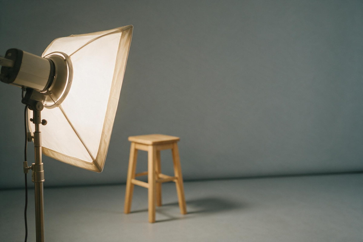
Light from above deepens the shadow in the tear trough and under the brow; light from the front fills it in. The same face can look tired in one and rested in the other with no surgery at all.
Tilting the chin down shows more upper lid; lifting it shows less. Camera distance and lens choice change facial proportions. Gaze direction changes how much white shows below the iris.
Raising the brows, even slightly, opens the eyes and mimics an upper lid result. Comparing a relaxed before with an alert after is not a comparison.
None of these has to be deliberate; they happen easily in a busy clinic. That is why standardised photography, same distance, same light, same position, same expression, is a marker of care in itself.
By six weeks a person looks socially recovered, which is what makes that time point popular. But healing is not finished. Fine swelling continues to resolve into the second and third month, the incision line is often at its pinkest around weeks three to four and only then begins to fade, and the lower lid position keeps settling as internal scar tissue matures. Scar maturation runs six to twelve months.
So an image labelled six weeks and an image labelled one year are answering different questions. If a gallery gives no time interval at all, that is a meaningful omission. The staged nature of recovery is set out in the day by day eyelid surgery recovery article.
For the upper lid the honest question is what created the change. Excess skin removal, the position of the crease and the level of the brow all contribute, and a photograph showing brighter eyes may reflect brow work rather than the eyelid operation. Comparing brow height between the two images is worth doing.
For the lower lid the questions are about contour and position. Look at the transition between lid and cheek, whether the lower lid margin sits at the same level in both images, and how much white shows beneath the iris. It is also worth knowing what surgery does not address: pigmentation of the skin under the eye is a different problem from a fat bulge and does not resolve because a bulge was corrected. The range of procedures involved is described on the cosmetic eyelid procedures page.
Some patterns should slow you down rather than reassure you:
A separate point: results that look unnaturally uniform across many different faces deserve scepticism, because faces are not uniform. Where an outcome has genuinely gone wrong, the issues involved are described in the article on unsatisfactory eyelid surgery.
A gallery becomes far more useful when you can interrogate it. Reasonable questions include: when was the after photograph taken; which technique was used; was anything else performed at the same session; are the photographs taken under standard conditions; can I see a case whose starting anatomy resembles mine; and what did that case look like at three months rather than at the end.
The things that determine your own outcome are not photographable. Skin quality and thickness, the strength and position of the lower lid, tear film and any tendency to dry eye, brow height, the amount and distribution of fat, previous surgery, thyroid eye disease, and the asymmetry that every face already has before anything is done. These are assessed by examination, and they matter more than any image on a screen.
My suggestion is to use galleries for one purpose only: to form questions. Bring them to an examination, look at your own anatomy in a mirror with someone who can explain what is technically possible for it, and make the decision from that conversation rather than from a photograph of somebody else.
They show what happened to another face with different anatomy, skin quality and starting point. They are useful for understanding the kind of change an operation produces, not for predicting your own outcome. A photograph cannot carry the information that decides your result.
There is no standard, which is exactly the problem. Images are taken anywhere from a few weeks to a year afterwards, and galleries often do not say. Without a time point beside the image, you cannot tell whether you are looking at a healing stage or a settled result.
No. At six weeks most visible swelling has gone, but fine oedema is still resolving, the incision line is still maturing and the lower lid position is still settling. Scar maturation runs six to twelve months, so a six-week image is an interim picture.
Overhead light deepens the shadows under the eyes and can make hollows look worse; frontal light flattens them. Chin position changes how much upper lid shows, and camera distance alters the proportions of the face. Change any of these between the two images and the difference is partly photographic.
Medical advertising rules in Turkey do not permit sharing patient images or case accounts in promotional material. This is why the article explains how to interpret galleries rather than showing any. Anatomy relevant to your own case can be discussed during an examination.
Consistent lighting, distance and head position between the two images, a stated time interval, several angles rather than one flattering view, similar expression and gaze direction, and clarity about which procedure was performed. Inconsistency in these is what makes a comparison unreliable.
Ask when the after image was taken, which technique was used, whether anything else was done at the same time, and whether you can see a case that started from anatomy similar to yours. The answers tell you more than the images do.
 English
