A blocked tear duct is an everyday problem in my clinic: constant watering, sticky discharge, and an eye that is never really comfortable. Many cases are managed without an operation. Some are not, and then the question becomes whether tear duct surgery, known as dacryocystorhinostomy or DCR, is the right step. This article explains when a blocked tear duct needs surgery, what DCR is in plain terms, how the external and endoscopic approaches differ, what happens before the operation, the first week afterwards, possible risks and failure, what it means when the duct blocks again, and when to see a lacrimal surgeon.
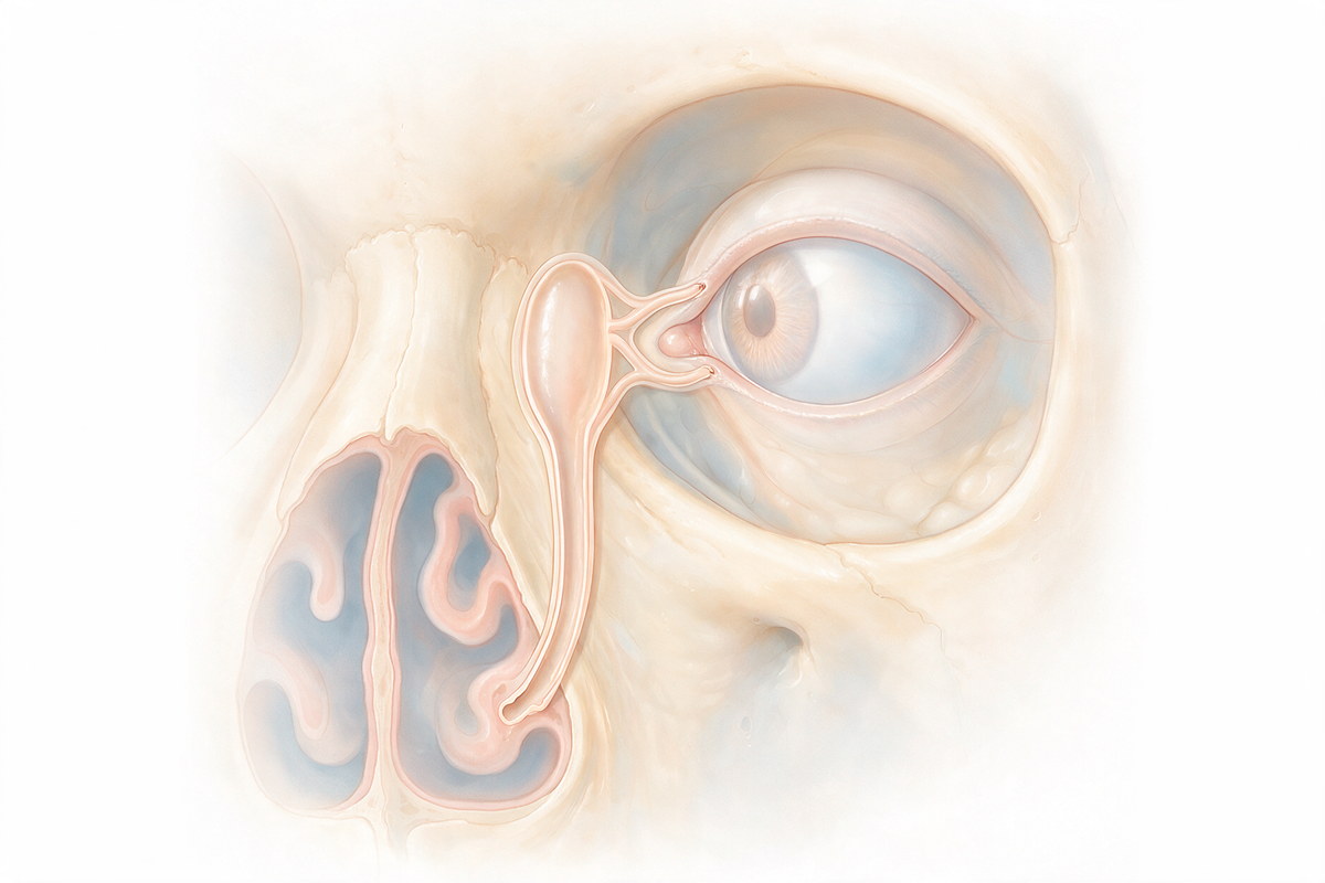
Tears drain through tiny openings in the inner corners of the lids, down into the tear sac, and along the nasolacrimal duct into the nose. When that duct is narrowed or blocked, tears cannot leave the eye. The result is watering that runs down the cheek, repeated sticky discharge, and sometimes infection of the tear sac. In adults, the blockage is usually a slow scarring narrowing with age and is rarely cured by drops or massage. Our page on adult nasolacrimal duct obstruction explains the causes in detail.
Surgery is considered when the watering is troublesome, when the blockage is confirmed on testing, and when infection of the tear sac keeps coming back. When an infection is acute, it is treated first. The decision depends on how much the symptoms affect your day, and it is made together with the surgeon.
DCR does not unblock the old duct. It creates a new passage. The surgeon makes a small opening in the thin bone between the tear sac and the inside of the nose, and the tear sac is connected directly to the nasal lining. Tears can then drain through the new route, bypassing the blockage. A small silicone tube is sometimes placed temporarily to keep the new opening open while it heals.
The operation is usually done under general anaesthesia, though some patients have it under local anaesthesia with sedation. Your surgeon will explain which is suitable.
There are two main approaches. In external DCR, a small cut is made on the skin beside the nose, and the connection is made through it. The scar usually fades well, and this approach gives the surgeon direct access. In endoscopic DCR, the surgeon works from inside the nose with a thin camera, so no skin cut is needed. Both are established methods, and both can be effective in the right patient.
The choice depends on the anatomy, previous surgery or infection, the surgeon's experience and your own preferences. Neither is the best choice for everyone, and a good surgeon explains the advantages and limits of each.
Before surgery, the lacrimal system is examined to confirm where the problem is. The tear passage is gently irrigated with saline to see where the fluid stops, and sometimes a scan or an endoscopic look at the nose is added. Problems inside the nose, such as a deviated septum or polyps, are looked for because they can affect the result. You will be asked about previous infection, injury, nasal surgery and any medicines that affect bleeding.
It helps to have realistic expectations. The aim is to improve watering and prevent infection, and many patients improve, but the outcome is not identical for everyone.
Mild bruising and some blood-stained fluid from the nose are common in the first days. The area can be swollen and tender. Your surgeon may advise nasal saline sprays and a short course of drops or medicines, and may ask you not to blow your nose forcefully for a period. Avoiding heavy lifting, bending and strenuous activity for a short time reduces bleeding. You will be told when to come for review and when the silicone tube, if one was placed, will be removed.
Watering usually improves over the following weeks as swelling settles, and your surgeon will check the opening during the follow-up visits.
All operations have risks. With DCR, they include bleeding from the nose, infection, scarring at the new opening, a visible skin scar after the external approach, and, occasionally, damage to nearby structures. The most important concern is that the new passage can narrow again. Failure is less common than success, but it does happen, and risk factors include scarring, chronic inflammation and anatomical problems in the nose. Your surgeon should explain these points before you consent.
Watering that returns weeks or months after surgery suggests that the new opening has closed or has become narrowed. The first step is a re-examination, because other causes of watering such as dry eye or lid problems can look similar. If the opening is blocked, revision surgery is possible, sometimes with a different technique or a tube. Our page on revision surgery for failed nasolacrimal duct obstruction treatment explains this in more detail.
Watering without redness, pain or swelling can be assessed at a routine appointment. Please arrange an examination if you have:
A red, hot, painful and swollen area at the inner corner of the eye with fever may be an infection of the tear sac that needs prompt treatment. Our page on dacryocystitis describes the condition. Spreading swelling around the eye, pain with eye movement or loss of vision is an emergency.
When watering is troublesome, the blockage is confirmed on testing, and drops or massage do not help, or when tear sac infections keep returning. In adults, a blockage from narrowing with age rarely clears without surgery.
Dacryocystorhinostomy creates a new drainage route by connecting the tear sac directly to the lining of the nose through a small opening in the bone. A small silicone tube is sometimes left briefly to keep it open.
In external DCR, a small skin cut is made beside the nose. In endoscopic DCR, the surgeon works from inside the nose with a thin camera and leaves no skin cut. Both are established, and the choice depends on the individual case.
Bruising and some bloody discharge from the nose are common for the first days. Many people return to light activities within a short period, but your surgeon will advise on timing and review visits.
Possible risks include bleeding, infection, scarring that narrows the new opening, a visible scar with the external approach and, rarely, damage to nearby structures. Your surgeon should explain them before consent.
Yes, the new opening can narrow or close again, especially with scarring or inflammation. Return of watering should be examined, and revision surgery is possible in suitable cases.
Mild or temporary blockages from infection or inflammation may improve, but a permanent narrowing of the duct in an adult seldom opens by itself. An examination confirms the cause and the options.
 English
