
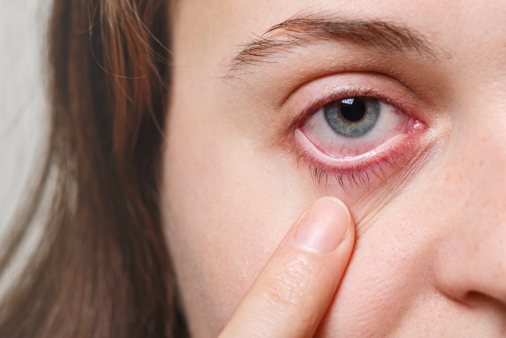
Eyelid tumors are abnormal growths that occur on the skin of the eyelids. These growths can be benign (non-cancerous) or malignant (cancerous), and they can affect both appearance and function. Left undiagnosed, they may lead to serious complications. Eyelid tumors are considered a form of skin cancer and are usually linked to sun exposure, genetic predisposition or environmental factors.
The eyelid carries the thinnest skin on the body and sits on one of the most sun-exposed parts of the face. Those two facts make the lid margin a common site for skin cancers. The reassuring part is that most lumps growing on the lid are benign, and an experienced ophthalmologist can distinguish between them in most patients at the examination, with the naked eye and under the microscope. Wherever doubt remains, a tissue sample is taken.
Patients who notice a lump on the lid often arrive saying they have a cyst. A cyst is a fluid-filled sac with clear borders, soft and generally harmless. It can arise from the sweat glands at the lid margin or from a blocked oil gland; small ones are simply watched, and those that cause discomfort are removed with a minor procedure.
The features that separate a cyst from a malignant lesion become clear at the examination. A cyst has smooth borders, does not break down the skin, does not cause lash loss, and the vessels over it look normal. In malignant lesions the borders become irregular, a central sore may form that will not heal, lashes fall out in that segment, the natural line of the lid margin is disrupted, and prominent vessels appear on the surface. I assess every lid lump against these criteria and send anything suspicious for biopsy. Congenital lesions form a separate topic, covered on the page about congenital masses and cysts, and vascular lesions on the hemangiomas page.
None of these findings means cancer on its own, but any lid lump that fails to settle within a few weeks, or that keeps growing, should be seen by an ophthalmologist.
The diagnosis is made by pathology. I therefore do not settle for saying a lesion looks benign; I seek a tissue diagnosis whenever there is doubt.
Small lesions can be removed under local anaesthesia in the office. Wide defects and repairs involving the lid margin require operating theatre conditions. Cold compresses reduce swelling in the first 48 hours, and sutures are removed during the first week depending on their position. Where cartilage grafts or flaps are used, the lid takes months to soften and settle into its final shape.
After a malignant tumor has been removed, follow-up is part of the treatment. Because a new lesion can appear in or near the same area, I see my patients more frequently during the first year and then at yearly intervals. I also recommend that patients with a history of skin cancer be assessed by a dermatologist for the rest of the face and body.
Eyelid tumors can be brought under control with early diagnosis and appropriate treatment. Because they can threaten eye health, it is important to consult an ophthalmologist as soon as symptoms are noticed. A lesion addressed early is removed with smaller surgery and the lid is easier to repair.
Ayşe Dolar Bilge MD, FEBOphth.
Ophthalmologist — Oculoplastic Surgery
As an Ophthalmologist, Dr. Ayşe Dolar Bilge, I provide diagnosis and treatment services with my experienced team.
Most lumps growing on the eyelid are benign, and an experienced ophthalmologist can distinguish between them in most patients at the examination, with the naked eye and under the microscope. Even so, any lid lump that fails to settle within a few weeks or keeps growing should be assessed, and suspicious ones biopsied.
A cyst is a fluid-filled sac with clear borders, soft and generally harmless. It can arise from the sweat glands at the lid margin or from a blocked oil gland. Small ones are simply watched; those causing discomfort are removed with a minor procedure. A cyst does not break down the skin, cause lash loss or disrupt the line of the lid margin.
The signs that raise suspicion are an irregularly bordered lump, a central sore that will not heal, loss of lashes over the area, disruption of the natural lid margin line, prominent surface vessels and growth over time. A chalazion-like swelling that keeps returning in the same place without responding to treatment should also be investigated.
A chalazion is a firm, painless nodule from a blocked oil gland; it usually settles with warm compresses and lid hygiene, whereas a tumor does not. A chalazion-like lesion that keeps recurring in the same place or fails to respond to treatment can suggest sebaceous carcinoma, a rare but aggressive tumor, and a tissue sample is taken.
Small lesions can be removed under local anaesthesia in the office. In malignant lesions a margin of healthy tissue is left and confirmed pathologically. Once the mass is removed the lid has to be repaired to restore both function and appearance; depending on the size of the defect this means direct closure, a flap of neighbouring tissue, or cartilage and skin grafts.
When incisions are placed in the natural lid crease or behind the lid, the scar is largely hidden. Where a flap or graft is used for a wide defect, the scar looks red and raised in the early weeks, then fades and softens over months. The lid takes several months to settle into its final shape, so I do not judge the result early.
The eyelid carries the thinnest skin on the body and sits on a sun-exposed part of the face, so years of ultraviolet exposure is the main risk factor. Fair skin, a previous history of skin cancer, conditions that suppress the immune system and genetic predisposition all increase the risk.
Recurrence is uncommon when the tumor is removed with a margin of healthy tissue, but it is not impossible. Because a new lesion can appear in or near the same area, follow-up is part of the treatment. I see patients more frequently during the first year and yearly afterwards, and recommend dermatological review for those with a history of skin cancer.
To get detailed information, send us your contact information and we will call you as soon as possible. Contact us now
 English
